Soleus

soleus -- 2016
Soleus

Pictured above is the soleus muscle. It stabilizes your ankle and pulls your heel bone towards the back of your lower leg (plantar flexes your ankle). It attaches to most of the length of the back of both your lower leg bones from very near your knee to just above your heel bone, moreso on the fibula bone (the smaller outside bone) than the tibia (the large weight-bearing bone in the lower leg). It also attaches to the achilles tendon from the inside (the gastrocnemius muscle attaches to the achilles tendon from the outside).

If this muscle locks up, all action invloving the ankle will be compromised. It is EXTREMELY LIKELY to be the source of your lower leg troubles. If you have issues with shin splints, plantar fasciitis, toe pains, or anything involving the lower leg or foot, make sure you address the soleus during your stretching and SMR efforts. You may need to work other areas, but do not skip the soleus!

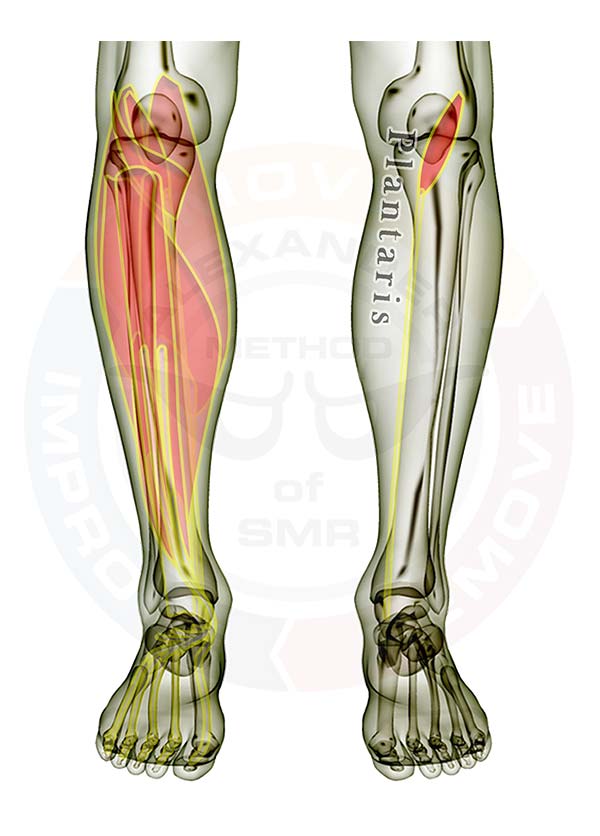
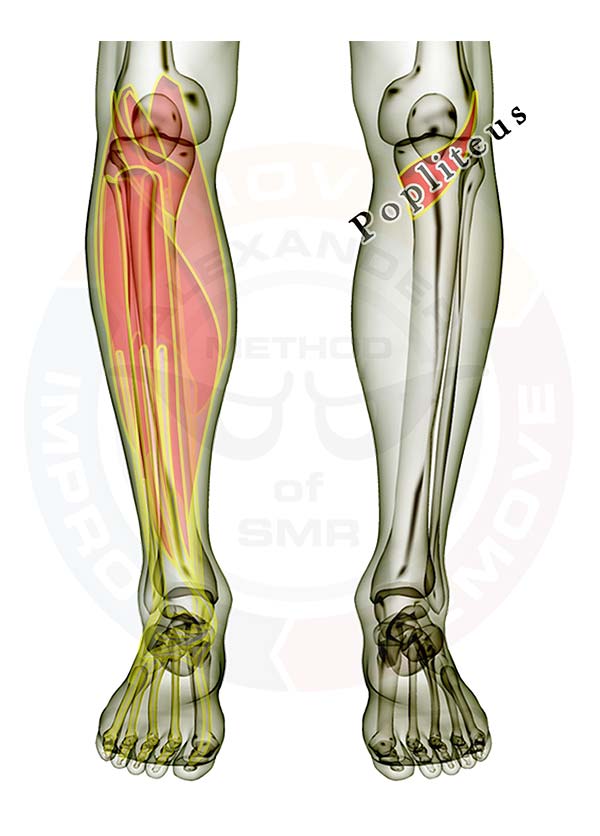
The soleus is displayed on the right leg. On the left leg the muscles are layered, showing how some of the muscles are covered by the others. All of the muscles are see-through so that you can appreciate the location and size of each muscle relative to the others.

Click here for a list of all the muscles.

Muscles that cross or attach to the hip or thigh bone (femur) and attach below the knee joint and DO NOT attach to the knee cap (patella)

  1. Sartorius
  2. Gracilis
  3. Biceps Femoris
  4. Semitendonosis
  5. Semimembranosis
  6. Gastrocnemius
  7. Popliteus
  8. Plantaris


Lower Leg
The following muscles attach across or below the knee.
Click the appropriate link for your interest.

Muscles that cross the knee

  1. Gastrocnemius
  2. Popliteus
  3. Plantaris


Muscles that pass or attach between the knee & ankle

  1. Gastrocnemius
  2. Popliteus
  3. Plantaris
  4. Soleus
  5. Peroneus Longus
  6. Peroneus Brevis
  7. Flexor Digitorum Longus
  8. Flexor Hallucis Longus
  9. Posterior Tibialis
  10. Anterior Tibialis
  11. Extensor Digitorum Longus
  12. Extensor Hallucis Longus


Good luck working out those tight knots.

If you have any questions, please post a comment. We try to respond within 24 hours.

We're here to help you get more out of your training!

Plantaris

Plantaris
Pictured above is the plantaris muscle. It helps bend your knee and raises your heel toward your knee. It attaches to the inside portion of the back of your thigh bone very near your knee and to the back of your heel bone. If this muscle locks up it will feel like a tight ball behind the knee. It is typically a symptom muscle instead of a source of your troubles. If you have plantaris issues you have OTHER bigger and stronger muscles that are not doing their job properly.

If this muscle regularly locks up on you, then you need to work on your quads EVERY DAY. Your quads extend (straighten) your knee, and the plantaris helps flex your knee. The quads are MUCH stronger than the plantaris (and all the other knee flexors, for that matter). In addition, your soleus muscle is probably locked up and your brain is firing the plantaris to help raise your heel more than it can handle. Be sure you check the soleus to relieve knots in it so your plantaris is no longer working harder than it should be.

The plantaris is displayed on the right leg. On the left leg the muscles are layered, showing how some of the muscles are covered by the others. All of the muscles are see-through so that you can appreciate the location and size of each muscle relative to the others.

Click here for a list of all the muscles.

Muscles that cross or attach to the hip or thigh bone (femur) and attach below the knee joint and DO NOT attach to the knee cap (patella)

  1. Sartorius
  2. Gracilis
  3. Biceps Femoris
  4. Semitendonosis
  5. Semimembranosis
  6. Gastrocnemius
  7. Popliteus
  8. Plantaris


Lower Leg
The following muscles attach across or below the knee.
Click the appropriate link for your interest.

Muscles that cross the knee

  1. Gastrocnemius
  2. Popliteus
  3. Plantaris


Muscles that pass or attach between the knee & ankle

  1. Gastrocnemius
  2. Popliteus
  3. Plantaris
  4. Soleus
  5. Peroneus Longus
  6. Peroneus Brevis
  7. Flexor Digitorum Longus
  8. Flexor Hallucis Longus
  9. Posterior Tibialis
  10. Anterior Tibialis
  11. Extensor Digitorum Longus
  12. Extensor Hallucis Longus


Good luck working out those tight knots.

If you have any questions, please post a comment. We try to respond within 24 hours.

We're here to help you get more out of your training!

Popliteus

Popliteus

Pictured above is the popliteus muscle. It unlocks and bends your knee. It attaches to the outer portion of the back of your thigh bone very near your knee and to the upper portion of the inside rear of your largest lower leg bone (the tibia). If this muscle locks up it will feel like a tight ball behind the knee.

If this muscle regularly locks up on you, then you need to work on your quads EVERY DAY. Your quads extend (straighten) your knee, and the popliteus flexes your knee. The quads are MUCH stronger than the popliteus (and all the other knee flexors, for that matter). Be sure you check the other muscles that attach to the knee when dealing with popliteus trouble.

The muscles are layered, showing how some of the muscles are covered by the others. All of the muscles are see-through so that you can appreciate the location and size of each muscle relative to the others.

Click here for a list of all the muscles.

Muscles that cross or attach to the hip or thigh bone (femur) and attach below the knee joint and DO NOT attach to the knee cap (patella)

  1. Sartorius
  2. Gracilis
  3. Biceps Femoris
  4. Semitendonosis
  5. Semimembranosis
  6. Gastrocnemius
  7. Popliteus
  8. Plantaris


Lower Leg
The following muscles attach across or below the knee.
Click the appropriate link for your interest.

Muscles that cross the knee

  1. Gastrocnemius
  2. Popliteus
  3. Plantaris


Muscles that pass or attach between the knee & ankle

  1. Gastrocnemius
  2. Popliteus
  3. Plantaris
  4. Soleus
  5. Peroneus Longus
  6. Peroneus Brevis
  7. Flexor Digitorum Longus
  8. Flexor Hallucis Longus
  9. Posterior Tibialis
  10. Anterior Tibialis
  11. Extensor Digitorum Longus
  12. Extensor Hallucis Longus


Good luck working out those tight knots.

If you have any questions, please post a comment. We try to respond within 24 hours.

We're here to help you get more out of your training!

Gastrocnemius

Gastrocnemius


Pictured above is the gastrocnemius muscle. It pulls your heel toward your thigh and also flexes your knee. It attaches to the back of your thigh bone very near your knee and to the upper portion of the outside of your Achilles tendon. Your soleus muscle also attaches to the Achilles tendon, but on the inside (the other side that the gastroc attaches to). Your soleus muscle is VERY likely to need LOTS of stretching and SMR attention. Occasionally, the gastroc will need attention too.

The muscles are layered, showing how some of the muscles are covered by the others. All of the muscles are see-through so that you can appreciate the location and size of each muscle relative to the others.

Click here for a list of all the muscles.

Muscles that cross or attach to the hip or thigh bone (femur) and attach below the knee joint and DO NOT attach to the knee cap (patella)

  1. Sartorius
  2. Gracilis
  3. Biceps Femoris
  4. Semitendonosis
  5. Semimembranosis
  6. Gastrocnemius
  7. Popliteus
  8. Plantaris


Lower Leg
The following muscles attach across or below the knee.
Click the appropriate link for your interest.

Muscles that cross the knee

  1. Gastrocnemius
  2. Popliteus
  3. Plantaris


Muscles that pass or attach between the knee & ankle

  1. Gastrocnemius
  2. Popliteus
  3. Plantaris
  4. Soleus
  5. Peroneus Longus
  6. Peroneus Brevis
  7. Flexor Digitorum Longus
  8. Flexor Hallucis Longus
  9. Posterior Tibialis
  10. Anterior Tibialis
  11. Extensor Digitorum Longus
  12. Extensor Hallucis Longus


Good luck working out those tight knots.

If you have any questions, please post a comment. We try to respond within 24 hours.

We're here to help you get more out of your training!

Foot Roll

Foot-Roll-1a--gallery
2nd Secondary -- Foot Roll
Use this SMR exercise to address the muscles in the bottom of your foot. If you have trouble with heel pain, fallen arches, claw/hammer toes, or just about any other discomfort or dysfunction in your foot, then this exercise is for you!

There are 11 muscles in the foot with 8 muscles on the bottom separated into 4 different layers. Start with light, slow movements. Address one foot at a time if necessary.

Gradually increase the pressure, but try not to roll quickly through the muscles in the bottom of your foot if you are interested in addressing the deeper layers (you'll be wasting your time).

Let your pain threshold guide you to back off when you apply too much pressure.

The best time to do this SMR exercise is just after a hot bath or shower, when your muscles are all warmed up.

You should do this movement at least once every 2 weeks. (more often is better)
For written instructions, click here to download a printable PDF of the Foot Roll

For our downloadable video instruction, click here (video from 2010--updated video is in the works!)

Be sure to post your questions and comments below. We want to provide the best instruction to help you recover from your workouts.

Click here to return to all of the Fundamental SMR Techniques

**All information is provided for educational purposes only. You should consult your doctor before attempting any exercises you read on this page or any page on this website.**

Calves Roll

calves-roll-1e--gallery
1st Secondary -- Calves Roll

Use this SMR exercise to address the muscles in the back of your calf.

If you have trouble with shin splints, heel pain or achilles tendon pain, or just about any other discomfort in your calves, then this exercise is for you!

The soleus muscle is involved in everything you do that involves the foot.

When it locks up, all foot movements (and even some knee & hip movements) are compromised.

You should do this movement at least once every 2 weeks. (more often is better)

For written instructions, click here to download a printable PDF of the Calves Roll

For our downloadable video instruction, click here (video from 2010--updated video is in the works!)

Be sure to post your questions and comments below. We want to provide the best instruction to help you recover from your workouts.

Click here to return to all of the Fundamental SMR Techniques

**All information is provided for educational purposes only. You should consult your doctor before attempting any exercises you read on this page or any page on this website.**