Pictured at left is the ISO light blue Rollga roller. It is "firm," but gives a little so that it isn't too hard for most people to work with.
The contours can be used to work out those muscular knots in your thighs, back, calves, and elsewhere MUCH better than with a traditional smooth roller.
Click here for an introductory video from Jeff and two of his Master Trainers.
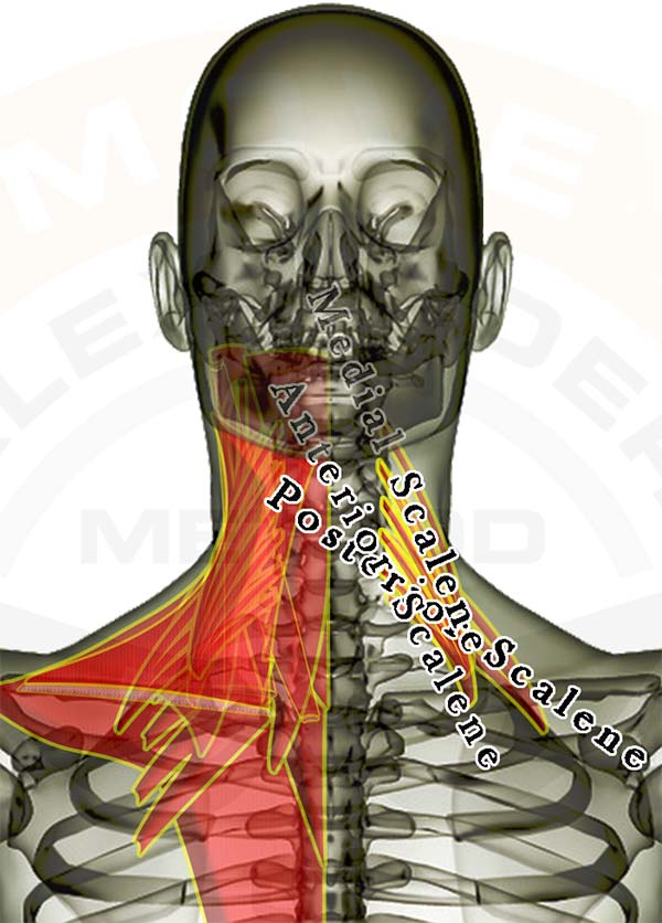
Pictured above are the scalene muscles. They lie underneath the sternocleidomastoid, originate along the front & side of your neck, and attach to the first and second ribs. They have a lot of influence on proper neck posture, and are typically "stress muscles."
The image of the muscles is layered, showing how some of the muscles are covered by the others. All of the muscles are see-through so that you can appreciate the location and size of each muscle relative to the others.
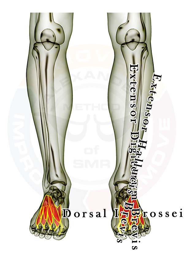
Muscles of the Foot
Pictured above are all the muscles in the top of the foot. As you can see there are MANY little muscles in this small area. It is best to approach the individual muscles initially as a group instead of starting with the individual muscles. If you find you need to go deeper to isolate one individual area or muscle then take the process in stages. Releasing tension in the entire foot can allow you to more easily reach the specific muscle you wish to address.
With any foot issue it is wise to continually address the largest muscles that insert into your foot, which are the toe and arch muscles that originate in the calves.
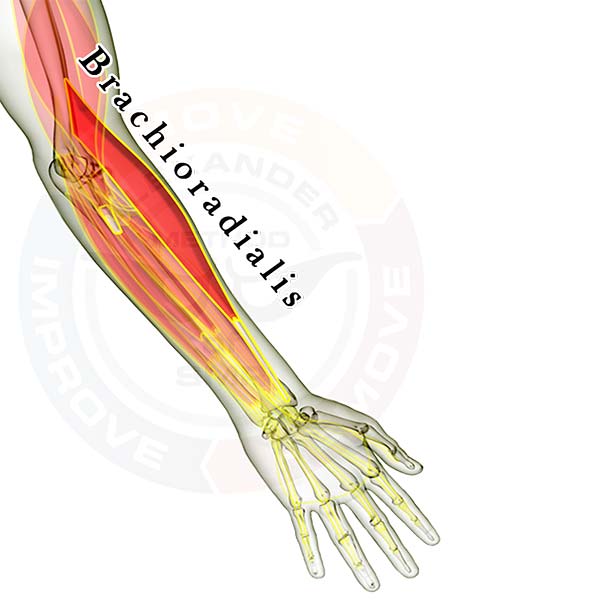
Pictured above is the brachioradialis muscle along with the other muscles located on the front of the forearm. It assists the biceps muscles by pulling your wrist toward your shoulder. It also helps stabilize the elbow during flexion or extension.
The muscles are layered, showing how some of the muscles are covered by the others. All of the muscles are see-through so that you can appreciate the location and size of each muscle relative to the others. You can access detail for all the muscles in the body with our Coach membership.
The following muscles cross the elbow joint and attach to the shoulder blade (scapula) & the arm (radius or ulna), to the upper arm (humerus) & the arm (radius or ulna). Click the appropriate link for your interest. Arm muscles that cross the elbow joint and flex the arm:
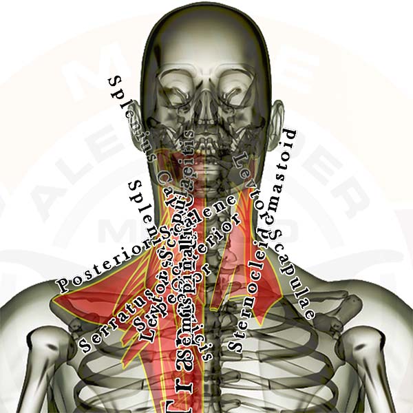
Pictured above are the muscles in the back of the neck. The levator scapulae and sternocleidomastoid are labelled on the right and all of the muscles in the back of the neck are listed on the left. The levator scapulae and sternocleidomastoid are very influential muscles for proper neck posture. Click Here for SMR technique instructions.
The muscles are layered, showing how some of the muscles are covered by others. All of the muscles are see-through so that you can appreciate the location and size of each muscle relative to the others.
Individual upper back & neck muscles you might be interested in: (any inactive links will be live soon)
Muscles that attach to the head (mastoid or other skull bone) and the neck (cervical vertebrae) and/or head & torso (thoracic vertebrae and/or ribs)
Listed below are all of the SMR Techniques, Anatomy, and any other posts on this site that you have selected as your favorites. This list should serve as your "go-to" areas to get your best self-care results.
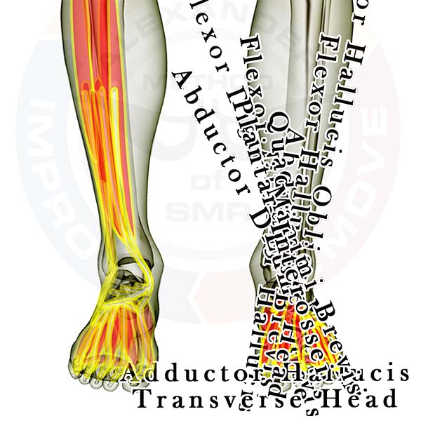
Muscles of the Foot
Pictured above are all the muscles in the bottom of the foot. As you can see there are MANY little muscles in the small area of your foot. It is best to approach the individual muscles initially as a group and shift attention to the forefoot, arch, or heel instead of starting with the individual muscles. If you find you need to go deeper to isolate one individual area or muscle take the process in stages. Releasing tension in the entire foot can allow you to more easily reach the specific muscle you wish to address.
With any foot issue it is wise to continually address the largest muscles that insert into your foot, the toe and arch muscles that originate in the calves.
This is likely to be the most productive adductor stretch you ever perform. When practiced correctly many people can see dramatic improvements in abduction of the hip in a fairly short period of time. There is one caveat: be sure you use your arms to bring your legs together following this stretch. Too many people are too aggressive when they exit a passive (static) stretch, and it is the aggressive exit that negatively impacts their athletic performance.
If you lengthen your muscles beyond their functional capacity to lift your own limbs (which this particular stretch can do to your Adductors), and then you lift your limbs immediately to get back to a normal position, you can injure the very muscles you just stretched. Remember the Stretch Reflex and learn how to feel your muscles "let go." When they do let go, assist them when exiting the stretch.
Some people perform a version of this stretch in which they pull on their leg to stretch their opposite hip. We suggest that you learn how to relax first before attempting to use force to encourage any muscle to relax. By using the wall as a support in this stretch and "blocking" the ankle across your knee it is possible to concentrate on melting into the floor with your lower back, completely relaxing your hip and thigh muscles, and getting much deeper stretch in your Piriformis and other lateral rotators.
For an older video of how to do the Wall Piriformis Block, click here. A new video is coming soon.
This stretch is to the chest and shoulders what the Supported Corpse is to the hips and lower back. Many people develop an anterior rotation of the shoulders or a shoulder-forward posture as they age. This is one way to reverse some of that shortening of the muscles that cross the front of the shoulders. Much like with the Chin Tuck or the Supported Corpse, the hardest part of this stretch for some people is doing nothing for more than 2 minutes. By constantly moving the overactive muscles of the shoulders never truly shut down. This means they take much longer to ever lengthen and it will be harder to note progress.
Breathe deeply and feel your knuckles melt into the floor. When you feel your elbows also melt into the floor you are ready to raise your arms a little closer to your ears.
Video coming soon.
Manage Cookie Consent
To provide the best experiences, we use technologies like cookies to store and/or access device information. Consenting to these technologies will allow us to process data such as browsing behavior or unique IDs on this site. Not consenting or withdrawing consent, may adversely affect certain features and functions.
Functional
Always active
The technical storage or access is strictly necessary for the legitimate purpose of enabling the use of a specific service explicitly requested by the subscriber or user, or for the sole purpose of carrying out the transmission of a communication over an electronic communications network.
Preferences
The technical storage or access is necessary for the legitimate purpose of storing preferences that are not requested by the subscriber or user.
Statistics
The technical storage or access that is used exclusively for statistical purposes.The technical storage or access that is used exclusively for anonymous statistical purposes. Without a subpoena, voluntary compliance on the part of your Internet Service Provider, or additional records from a third party, information stored or retrieved for this purpose alone cannot usually be used to identify you.
Marketing
The technical storage or access is required to create user profiles to send advertising, or to track the user on a website or across several websites for similar marketing purposes.
 Pictured at left is the ISO light blue Rollga roller. It is "firm," but gives a little so that it isn't too hard for most people to work with.