Pictured above is the tensor fasciae latae. It primarily acts on the IT Band and keeps the knee from drifting inward. It is also a weak hip flexor muscle. You can't move your hip in any direction without using this muscle. Cyclists, speed skaters and other athletes that spend large amounts of time actively using their hip in a flexed position are likely to need stretching and SMR attention here.
80% of all adults over 18 years of age have some form of back pain. If you aren't also checking the psoas and rectus femoris for excessive tension you are missing the most significant inhibitors of proper back function.
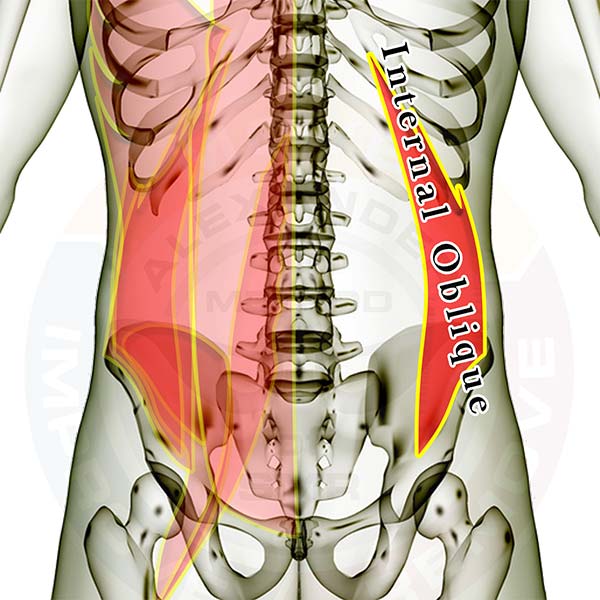
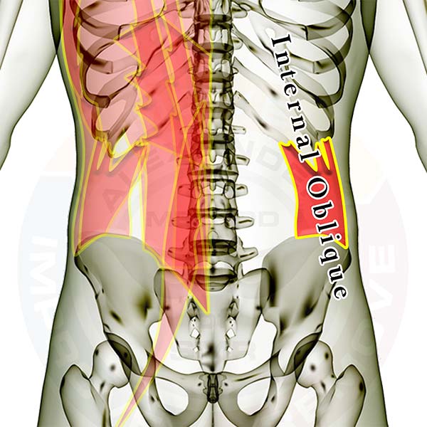
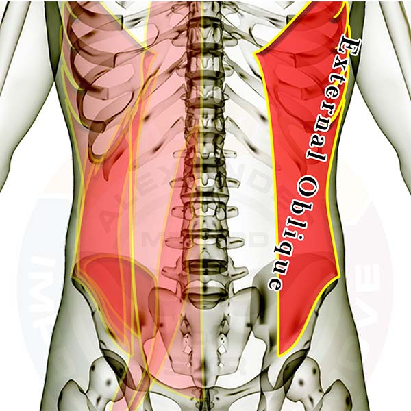
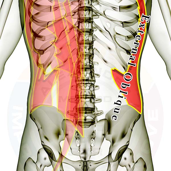
The muscles are layered, showing how some of the muscles are covered by the others. All of the muscles are see-through so that you can appreciate the location and size of each muscle relative to the others.
Individual hip, lower back & thigh muscles you might be interested in: (any inactive links will be live soon)
Click here for a list of all the muscles.
Click here to see the hip flexor muscles as a group.
Muscle that crosses the hip/lower back joint and crosses the hip/thigh joint (attaches to the spine and the femur)
Muscles that attach to the hip and the spine and/or ribs
- Rectus Abdominus
- External Abdominal Oblique
- Internal Abdominal Oblique
- Transverse Abdominus
- Latissimus Dorsi
- Iliocostalis Lumborum
- Longissimus Thoracis
- Quadratus Lumborum
- Multifidi
Muscles that attach to the hip and the thigh bone (femur)
- Iliacus
- Rectus Femoris
- Tensor Fasciae Latae
- Sartorius
- Gracilis
- Adductor Magnus
- Adductor Longus
- Adductor Brevis
- Pectineus
- Gluteus Maximus
- Gluteus Medius
- Gluteus Minimus
- Piriformis
- Superior Gemellus
- Obturator Internus
- Inferior Gemellus
- Obturator Externus
- Quadratus Femoris
- Biceps Femoris
- Semitendonosis
- Semimembranosis
Good luck working out those tight knots.
If you have any questions, please post a comment. We try to respond within 24 hours.
We're here to help you get more out of your training!