Scalenes

scalenes -- 2016
Scalenes

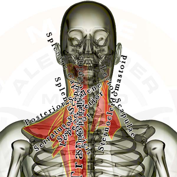
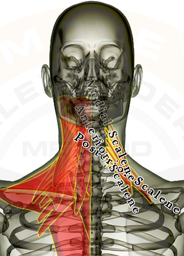
Pictured above are the scalene muscles. They lie underneath the sternocleidomastoid, originate along the front & side of your neck, and attach to the first and second ribs. They have a lot of influence on proper neck posture, and are typically "stress muscles."

The image of the muscles is layered, showing how some of the muscles are covered by the others. All of the muscles are see-through so that you can appreciate the location and size of each muscle relative to the others.

Click here for a list of all the muscles.

Individual upper back & neck muscles you might be interested in: (any inactive links will be live soon)

Muscles that attach to the head (mastoid or other skull bone) and the neck (cervical vertebrae) and/or head & torso (thoracic vertebrae and/or ribs)

  1. Sternocleidomastoid
  2. Trapezius
  3. Splenius Capitus
  4. Longissimus Capitus
  5. Semispinalis Capitus

Muscles that attach to the neck (cervical vertebrae) and another area of the spine (cervical or thoracic vertebrae) and/or ribs

  1. Anterior Scalene
  2. Medial Scalene
  3. Posterior Scalene
  4. Splenius Cervicis
  5. Semispinalis Cervicis

Muscles that attach to the head or neck & the shoulder blade (scapula)

  1. Trapezius
  2. Levator Scapulae

Good luck working out those tight knots.

If you have any questions, please post a comment. We try to respond within 24 hours.

We're here to help you get more out of your training!

Top of Foot

foot -- 2016
Muscles of the Foot

Pictured above are all the muscles in the top of the foot. As you can see there are MANY little muscles in this small area. It is best to approach the individual muscles initially as a group instead of starting with the individual muscles. If you find you need to go deeper to isolate one individual area or muscle then take the process in stages. Releasing tension in the entire foot can allow you to more easily reach the specific muscle you wish to address.

With any foot issue it is wise to continually address the largest muscles that insert into your foot, which are the toe and arch muscles that originate in the calves.

Click here for a list of all the muscles.


Lower Leg
The following muscles attach across or below the knee. Click the appropriate link for your interest.

Muscles that cross the knee

  1. Gastrocnemius
  2. Popliteus
  3. Plantaris


Muscles that pass or attach between the knee & ankle

  1. Gastrocnemius
  2. Popliteus
  3. Plantaris
  4. Soleus
  5. Peroneus Longus
  6. Peroneus Brevis
  7. Flexor Digitorum Longus
  8. Flexor Hallucis Longus
  9. Posterior Tibialis
  10. Anterior Tibialis
  11. Extensor Digitorum Longus
  12. Extensor Hallucis Longus


Muscles that cross the ankle

  1. Flexor Digitorum Longus
  2. Flexor Hallucis Longus
  3. Posterior Tibialis
  4. Peroneus Longus
  5. Peroneus Brevis
  6. Anterior Tibialis
  7. Extensor Digitorum Longus
  8. Extensor Hallucis Longus


Muscles in the bottom of the foot (active links coming soon)

  1. Flexor Digitorum Brevis
  2. Abductor Hallucis
  3. Abductor Digiti Minimi
  4. Flexor Hallucis Brevis
  5. Quadratus Planatae
  6. Lumbricals
  7. Adductor Hallucis
  8. Flexor Digitorum Minimi Brevis
  9. Plantar Interossei


Muscles in the top of the foot (active links coming soon)

  1. Extensor Digitorum Brevis
  2. Extensor Hallucis Brevis
  3. Dorsal Interossei


Good luck working out those tight knots.

If you have any questions, please post a comment. We try to respond within 24 hours.

We're here to help you get more out of your training!

Wrist Extensors

brachioradialis -- 2016
Brachioradialis - Front


Pictured above is the brachioradialis muscle along with the other muscles located on the front of the forearm. It assists the biceps muscles by pulling your wrist toward your shoulder. It also helps stabilize the elbow during flexion or extension.

The muscles are layered, showing how some of the muscles are covered by the others. All of the muscles are see-through so that you can appreciate the location and size of each muscle relative to the others. You can access detail for all the muscles in the body with our Coach membership.

Click here for a list of all the muscles.

Click here to see the biceps muscles as a group.

The following muscles cross the elbow joint and attach to the shoulder blade (scapula) & the arm (radius or ulna), to the upper arm (humerus) & the arm (radius or ulna). Click the appropriate link for your interest.

Arm muscles that cross the elbow joint and flex the arm:

  1. Biceps Brachii
  2. Brachialis
  3. Brachioradialis - Front


Good luck working out those tight knots.

If you have any questions, please post a comment. We try to respond within 24 hours.

We're here to help you get more out of your training!

Neck — Back

neck -- 2016

Pictured above are the muscles in the back of the neck. The levator scapulae and sternocleidomastoid are labelled on the right and all of the muscles in the back of the neck are listed on the left. The levator scapulae and sternocleidomastoid are very influential muscles for proper neck posture. Click Here for SMR technique instructions.

Click Here for a list of all the muscles.

The muscles are layered, showing how some of the muscles are covered by others. All of the muscles are see-through so that you can appreciate the location and size of each muscle relative to the others.

Individual upper back & neck muscles you might be interested in: (any inactive links will be live soon)

Muscles that attach to the head (mastoid or other skull bone) and the neck (cervical vertebrae) and/or head & torso (thoracic vertebrae and/or ribs)

  1. Sternocleidomastoid
  2. Trapezius
  3. Splenius Capitus
  4. Longissimus Capitus
  5. Semispinalis Capitus

Muscles that attach to the neck (cervical vertebrae) and another area of the spine (cervical or thoracic vertebrae) and/or ribs

  1. Anterior Scalene
  2. Medial Scalene
  3. Posterior Scalene
  4. Splenius Cervicis
  5. Semispinalis Cervicis

Muscles that attach to the head or neck & the shoulder blade (scapula)

  1. Trapezius
  2. Levator Scapulae

Good luck working out those tight knots.

If you have any questions, please post a comment. We try to respond within 24 hours.

We're here to help you get more out of your training!

Foot

foot -- 2016
Muscles of the Foot

Pictured above are all the muscles in the bottom of the foot. As you can see there are MANY little muscles in the small area of your foot. It is best to approach the individual muscles initially as a group and shift attention to the forefoot, arch, or heel instead of starting with the individual muscles. If you find you need to go deeper to isolate one individual area or muscle take the process in stages. Releasing tension in the entire foot can allow you to more easily reach the specific muscle you wish to address.

With any foot issue it is wise to continually address the largest muscles that insert into your foot, the toe and arch muscles that originate in the calves.

Click here for a list of all the muscles.


Lower Leg
The following muscles attach across or below the knee. Click the appropriate link for your interest.

Muscles that cross the knee

  1. Gastrocnemius
  2. Popliteus
  3. Plantaris


Muscles that pass or attach between the knee & ankle

  1. Gastrocnemius
  2. Popliteus
  3. Plantaris
  4. Soleus
  5. Peroneus Longus
  6. Peroneus Brevis
  7. Flexor Digitorum Longus
  8. Flexor Hallucis Longus
  9. Posterior Tibialis
  10. Anterior Tibialis
  11. Extensor Digitorum Longus
  12. Extensor Hallucis Longus


Muscles that cross the ankle

  1. Flexor Digitorum Longus
  2. Flexor Hallucis Longus
  3. Posterior Tibialis
  4. Peroneus Longus
  5. Peroneus Brevis
  6. Anterior Tibialis
  7. Extensor Digitorum Longus
  8. Extensor Hallucis Longus


Muscles in the bottom of the foot (active links coming soon)

  1. Flexor Digitorum Brevis
  2. Abductor Hallucis
  3. Abductor Digiti Minimi
  4. Flexor Hallucis Brevis
  5. Quadratus Planatae
  6. Lumbricals
  7. Adductor Hallucis
  8. Flexor Digitorum Minimi Brevis
  9. Plantar Interossei


Muscles in the top of the foot (active links coming soon)

  1. Extensor Digitorum Brevis
  2. Extensor Hallucis Brevis
  3. Dorsal Interossei


Good luck working out those tight knots.

If you have any questions, please post a comment. We try to respond within 24 hours.

We're here to help you get more out of your training!

Serratus Posterior Inferior

spi -- 2016
Serratus Posterior Inferior

Pictured above is the serratus posterior inferior (SPI). It is an important muscle to address because it tends to be tight and limit your ability to draw in a deep breath.

It is commonly overstretched and lacking proper mobility in persons with poor posture due to excessive rounding of the middle and lower back. 80% of all adults over 18 years of age have some form of back pain. If you aren't checking the SPI for excessive stiffness you are missing one of the most significant links to proper back function.

The muscles on the left are layered, showing how some of the muscles are covered by the others. All of the muscles are see-through so that you can appreciate the location and size of each muscle relative to the others.

Click here for a list of all the muscles.

Individual hip, lower back & thigh muscles you might be interested in: (any inactive links will be live soon)

The following muscle attaches across the middle & lower back to the bottom four ribs and the last two thoracic vertebrae and the top two lumbar vertebrae:

  1. Serratus Posterior Inferior

The following muscles attach across the hip & lower back, or to the hip from above (the spine and/or ribs) or below (the thigh).
Click the appropriate link for your interest.

Click here to see the hip flexors as a muscle group.

Click here to see the adductors as a group.

Muscle that crosses the hip/lower back joint and crosses the hip/thigh joint. (attaches to the spine and the femur)

  1. Psoas


Muscles that attach to the hip and the spine and/or ribs

  1. Rectus Abdominus
  2. External Abdominal Oblique
  3. Internal Abdominal Oblique
  4. Transverse Abdominus
  5. Latissimus Dorsi
  6. Iliocostalis Lumborum
  7. Longissimus Thoracis
  8. Quadratus Lumborum
  9. Multifidi


Muscles that attach to the hip and the thigh bone (femur)

  1. Iliacus
  2. Adductor Magnus
  3. Adductor Longus
  4. Adductor Brevis
  5. Pectineus
  6. Gluteus Maximus
  7. Gluteus Medius
  8. Gluteus Minimus
  9. Piriformis
  10. Superior Gemellus
  11. Obturator Internus
  12. Inferior Gemellus
  13. Obturator Externus
  14. Quadratus Femoris

Good luck working out those tight knots.

If you have any questions, please post a comment. We try to respond within 24 hours.

We're here to help you get more out of your training!

Posterior Tibialis

Posterior Tibialis

Pictured above is the posterior tibialis muscle. It stabilizes your foot & ankle for balance by pulling the bones of your arch toward the back of your lower leg (plantar flexing the ankle) and rotating the bottom of your foot inward (inverting your foot). It attaches underneath all of the muscles attached to the back of your lower leg between the two bones, and its tendon runs down the back inside of your ankle and attaches to most of the bones in the arch of the foot.

If this muscle locks up it will feel tight and possibly painful in the bottom of your foot. This muscle is a BIG contributor plantar fasciitis. Rarely will this muscle lock up without the toe flexor muscles needing attention too. The soleus muscle ALWAYS needs attention if your posterior tibialis muscle locks up, and your peroneal muscles will also likely need some TLC on the outside of your lower leg.

The posterior tibialis muscle is displayed on the right leg. On the left leg the muscles located in the back of your lower leg are layered, showing how some of the muscles are covered by the others. All of the muscles are see-through so that you can appreciate the location and size of each muscle relative to the others.

Click here for a list of all the muscles.

Muscles that cross or attach to the hip or thigh bone (femur) and attach below the knee joint and DO NOT attach to the knee cap (patella)

  1. Sartorius
  2. Gracilis
  3. Biceps Femoris
  4. Semitendonosis
  5. Semimembranosis
  6. Gastrocnemius
  7. Popliteus
  8. Plantaris


Lower Leg
The following muscles attach across or below the knee.
Click the appropriate link for your interest.

Muscles that cross the knee

  1. Gastrocnemius
  2. Popliteus
  3. Plantaris


Muscles that pass or attach between the knee & ankle

  1. Gastrocnemius
  2. Popliteus
  3. Plantaris
  4. Soleus
  5. Peroneus Longus
  6. Peroneus Brevis
  7. Flexor Digitorum Longus
  8. Flexor Hallucis Longus
  9. Posterior Tibialis
  10. Anterior Tibialis
  11. Extensor Digitorum Longus
  12. Extensor Hallucis Longus


Good luck working out those tight knots.

If you have any questions, please post a comment. We try to respond within 24 hours.

We're here to help you get more out of your training!

Flexor Digitorum Longus

Flexor Digitorum Longus

Pictured above is the flexor digitorum longus muscle. It stabilizes your foot & ankle for balance by pulling all of your toes except the big toe toward the bottom of your foot. If contracted hard enough, it pulls the bottom of your foot toward the back of your lower leg like when you point your toes. It attaches underneath the soleus muscle along most of the back of the bigger of your lower leg bones (the tibia), and its tendon runs down the back inside of your ankle and attaches to the bottom of all the toes except the big toe.

If this muscle locks up it will feel tight and possibly painful along the center portion of the back of your lower leg, and your toes will not move well. This muscle and the flexor hallucis longus are BIG contributors to hammer toes, claw toes, and other general lower leg and foot pains. Rarely will this muscle lock up without the other toe flexor muscle needing attention too (the flexor hallucis longus). The soleus muscle ALWAYS needs attention if your flexor muscles are locked up, and your toe extensors will also likely need some TLC on the front of your lower leg deeper than the anterior tibialis.

The flexor digitorum longus muscle is displayed on the right leg. On the left leg the muscles located in the back of your lower leg are layered, showing how some of the muscles are covered by the others. All of the muscles are see-through so that you can appreciate the location and size of each muscle relative to the others. You can access detail for all the muscles in the body with our Coach membership.

Click here for a list of all the muscles.

Muscles that cross or attach to the hip or thigh bone (femur) and attach below the knee joint and DO NOT attach to the knee cap (patella)

  1. Sartorius
  2. Gracilis
  3. Biceps Femoris
  4. Semitendonosis
  5. Semimembranosis
  6. Gastrocnemius
  7. Popliteus
  8. Plantaris


Lower Leg
The following muscles attach across or below the knee.
Click the appropriate link for your interest.

Muscles that cross the knee

  1. Gastrocnemius
  2. Popliteus
  3. Plantaris


Muscles that pass or attach between the knee & ankle

  1. Gastrocnemius
  2. Popliteus
  3. Plantaris
  4. Soleus
  5. Peroneus Longus
  6. Peroneus Brevis
  7. Flexor Digitorum Longus
  8. Flexor Hallucis Longus
  9. Posterior Tibialis
  10. Anterior Tibialis
  11. Extensor Digitorum Longus
  12. Extensor Hallucis Longus


Good luck working out those tight knots.

If you have any questions, please post a comment. We try to respond within 24 hours.

We're here to help you get more out of your training!

Flexor Hallucis Longus

Flexor Hallucis Longus

Pictured above is the flexor hallucis longus muscle (the 'big toe flexor'). It stabilizes your foot & ankle for balance by pulling your big toe toward the bottom of your foot. If contracted hard enough, it pulls the bottom of your foot toward the back of your lower leg like when you point your toes. It attaches underneath the soleus muscle on the lower 2/3 of the back of your lower leg bones moreso on the smaller outer bone (the fibula), and its tendon runs down the back inside of your ankle and attaches to the bottom of only the big toe.

If this muscle locks up it will feel tight and possibly painful along the lower portion of the back of your lower leg, and your big toe will not move well. This muscle and the flexor digitorum longus are BIG contributors to hammer toes, claw toes, and other general lower leg and foot pains. Rarely will this muscle lock up without the other toe flexor muscle needing attention too (the flexor digitorum longus). The soleus muscle ALWAYS needs attention if your flexor muscles are locked up, and your toe extensors will also likely need some TLC on the front of your lower leg deeper than the anterior tibialis.

The flexor hallucis longus muscle is displayed on the right leg. On the left leg the muscles located in the back of your lower leg are layered, showing how some of the muscles are covered by the others. All of the muscles are see-through so that you can appreciate the location and size of each muscle relative to the others. You can access detail for all the muscles in the body with our Coach membership.

Click here for a list of all the muscles.

Muscles that cross or attach to the hip or thigh bone (femur) and attach below the knee joint and DO NOT attach to the knee cap (patella)

  1. Sartorius
  2. Gracilis
  3. Biceps Femoris
  4. Semitendonosis
  5. Semimembranosis
  6. Gastrocnemius
  7. Popliteus
  8. Plantaris


Lower Leg
The following muscles attach across or below the knee.
Click the appropriate link for your interest.

Muscles that cross the knee

  1. Gastrocnemius
  2. Popliteus
  3. Plantaris


Muscles that pass or attach between the knee & ankle

  1. Gastrocnemius
  2. Popliteus
  3. Plantaris
  4. Soleus
  5. Peroneus Longus
  6. Peroneus Brevis
  7. Flexor Digitorum Longus
  8. Flexor Hallucis Longus
  9. Posterior Tibialis
  10. Anterior Tibialis
  11. Extensor Digitorum Longus
  12. Extensor Hallucis Longus


Good luck working out those tight knots.

If you have any questions, please post a comment. We try to respond within 24 hours.

We're here to help you get more out of your training!

Extensor Hallucis Longus

ext hal long -- 2016
Extensor Hallucis Longus

Pictured above is the extensor hallucis longus muscle (the 'big toe muscle'). It stabilizes your foot & ankle for balance by pulling your big toe toward the front of your shin. It attaches underneath the anterior tibialis to the lower half of the front of your lower leg bone, and its tendon runs down the front of your ankle and attaches to only the big toe.

If this muscle locks up it will feel tight and possibly painful along the lower portion of the front of your lower leg, and your big toe will not move well. This muscle and the extensor digitorum longus are BIG contributors to shin splints, hammer toes, claw toes, and other shin and foot pains. Rarely will this muscle lock up without the other toe extensor muscle needing attention too (the extensor digitorum longus). The soleus muscle ALWAYS needs attention if your extensor muscles are locked up, and your toe flexors will also likely need some TLC on the back of your lower leg deeper than the soleus.

The extensor hallucis longus muscle is displayed on the right leg. On the left leg the muscles located in the front of your shin are layered, showing how some of the muscles are covered by the others. All of the muscles are see-through so that you can appreciate the location and size of each muscle relative to the others.

Click here for a list of all the muscles.

Click here to see all of the foot muscles.
Muscles that cross or attach to the hip or thigh bone (femur) and attach below the knee joint and DO NOT attach to the knee cap (patella)

  1. Sartorius
  2. Gracilis
  3. Biceps Femoris
  4. Semitendonosis
  5. Semimembranosis
  6. Gastrocnemius
  7. Popliteus
  8. Plantaris


Lower Leg
The following muscles attach across or below the knee.
Click the appropriate link for your interest.

Muscles that cross the knee

  1. Gastrocnemius
  2. Popliteus
  3. Plantaris


Muscles that pass or attach between the knee & ankle

  1. Gastrocnemius
  2. Popliteus
  3. Plantaris
  4. Soleus
  5. Peroneus Longus
  6. Peroneus Brevis
  7. Flexor Digitorum Longus
  8. Flexor Hallucis Longus
  9. Posterior Tibialis
  10. Anterior Tibialis
  11. Extensor Digitorum Longus
  12. Extensor Hallucis Longus


Good luck working out those tight knots.

If you have any questions, please post a comment. We try to respond within 24 hours.

We're here to help you get more out of your training!